Today in class, my partner and I dissected a sheep eye to better understand the structure and physiology of the eye. A sheep eye is a little bit different from the human eye, for example, a sheep's pupil is oblong shaped, while the human eye is circular. Additionally, the sheep eye has a glittery layer behind the retina called the tapetum lucidum, which helps sheep see in the dark. Humans don't need this because we don't have night-vision.
The eye untouched:
We started the dissection by cutting away the fatty tissue, the muscle, and eye-lid from both the front and rear surfaces of the eye. Then we were left with an eye that looks like this:


Then we made an incision down the midway of the sclera between the cornea and the optic nerve.
This cut the eye in two anterior and posterior hemispheres. Each side looked like this:

On the posterior (back) hemisphere of the eye there is the retina when peeled to the side reveals a black underlying choroid coat. The retina remains attached in one place called the blind spot. Then we scraped away the choroid coat, which revealed a blue glittery layer called the tapetum lucidum, which, as I have said above, is not found in the human eye because. It reflect light onto the retina which helps certain animals see in the dark.


On the anterior hemisphere of the eye, there is the semi-fluid vitreous humor that fills the central cavity of the eye and the aqueous humor, which is found behind the cornea.
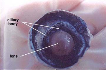
We scoped out the vitreous humor and the lens, this reveals the lens, ciliary body, and suspensory ligaments. The normal lens is convex shaped and somewhat elastic. It is held in place by the suspensory ligaments that in turn join with the smooth muscle containing ciliary body. We then removed the lens by pulling it free from its attachments. The lens was circular and felt kind of like uncooked tapioca.


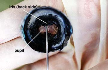
After removing the vitreous humor and the lens, we could now see an opening that allows light to enter the eye, the pupil, which is located in the center of the black iris. A sheep has an oblong- shaped pupil, while a human has a circular pupil. We then removed a thin filament protein covering the pupil on the front side of the hemisphere, called the cornea.


The eye untouched:
We started the dissection by cutting away the fatty tissue, the muscle, and eye-lid from both the front and rear surfaces of the eye. Then we were left with an eye that looks like this:
Then we made an incision down the midway of the sclera between the cornea and the optic nerve.
This cut the eye in two anterior and posterior hemispheres. Each side looked like this:
On the posterior (back) hemisphere of the eye there is the retina when peeled to the side reveals a black underlying choroid coat. The retina remains attached in one place called the blind spot. Then we scraped away the choroid coat, which revealed a blue glittery layer called the tapetum lucidum, which, as I have said above, is not found in the human eye because. It reflect light onto the retina which helps certain animals see in the dark.


On the anterior hemisphere of the eye, there is the semi-fluid vitreous humor that fills the central cavity of the eye and the aqueous humor, which is found behind the cornea.
We scoped out the vitreous humor and the lens, this reveals the lens, ciliary body, and suspensory ligaments. The normal lens is convex shaped and somewhat elastic. It is held in place by the suspensory ligaments that in turn join with the smooth muscle containing ciliary body. We then removed the lens by pulling it free from its attachments. The lens was circular and felt kind of like uncooked tapioca.


After removing the vitreous humor and the lens, we could now see an opening that allows light to enter the eye, the pupil, which is located in the center of the black iris. A sheep has an oblong- shaped pupil, while a human has a circular pupil. We then removed a thin filament protein covering the pupil on the front side of the hemisphere, called the cornea.


No comments:
Post a Comment