Saturday, April 30, 2016

Sheep Brain Dissection

Today in class, we dissected a brain. We only made three cuts but we identified most of the important structures of the brain, by using pins.
 Anterior (White) and Posterior (Black)
Cerebrum (yellow), Cerebellum (green), and Brain Stem (Red)


First we observed the external structure of the brain, starting with the meninges, the a shiny membrane substance surrounding the brain. After removing the meninges, we identified the anterior and posterior sides of the brain. Then we identified the cerebrum, cerebellum, and brain stem.


Cerebrum
is divided into four lobes and is responsible for body orientation, processing sensory input, cognition, memory, voluntary motor action, visual reception and association, auditory reception and interpretation, and expression powers.
Cerebellum
is responsible for motor control and coordination.
Brain Stem
performs necessary functions, like heart rate and breathing

Myelin is a mixture of proteins and phospholipids forming a whitish insulating sheath around many nerve fibers, increasing the speed at which impulses are conducted. 


Then we cut the brain longitudinally (like a hot dog) so that we could observe the medial plane of the brain. From this view of the brain we could see the white and grey matter of the brain. 

 Thalamus (yellow), Midbrain (blue), Medulla Oblongata(Red), Pons (Green)


 Hypothalamus (Blue)


Corpus Callosum (Red)                                                                                          Optic Nerve (Green)


Thalamus
works to correlate several important processes, including consciousness, sleep, and sensory interpretation.
Optic Nerve
transfer visual information from the retina to the vision centers of the brain via electrical impulses. The optic nerve is made of ganglionic cells or nerve cells.
Medulla Oblongata
regulate breathing, heart and blood vessel function, digestion, sneezing, and swallowing. This part of the brain is a center for respiration and circulation.
Pons
a bridge between various parts of the nervous system, including the cerebellum and cerebrum
Midbrain
is a portion of the central nervous system associated with vision, hearing, motor control, sleep/wake, arousal (alertness), and temperature regulation.
Corpus Callosum
Large bundle of neuron fibers (myelinated axons or white matter) connecting 2 hemispheres
Hypothalamus
Maintains homeostasis and links the nervous system to the endocrine system

Then we made a cross sectional cut of the cerebrum (like a hamburger).

White and Gray Matter


Wednesday, April 27, 2016

Sheep Eye Dissection

Today in class, my partner and I dissected a sheep eye to better understand the structure and physiology of the eye. A sheep eye is a little bit different from the human eye, for example, a sheep's pupil is oblong shaped, while the human eye is circular. Additionally, the sheep eye has a glittery layer behind the retina called the tapetum lucidum, which helps sheep see in the dark. Humans don't need this because we don't have night-vision.

The eye untouched:


We started the dissection by cutting away the fatty tissue, the muscle, and eye-lid from both the front and rear surfaces of the eye. Then we were left with an eye that looks like this:


Then we made an incision down the midway of the sclera between the cornea and the optic nerve.
This cut the eye in two anterior and posterior hemispheres. Each side looked like this:


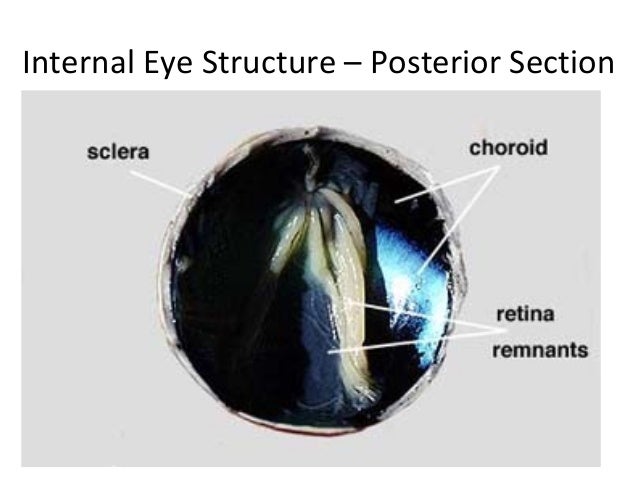
On the posterior (back) hemisphere of the eye there is the retina when peeled to the side reveals a black underlying choroid coat. The retina remains attached in one place called the blind spot. Then we scraped away the choroid coat, which revealed a blue glittery layer called the tapetum lucidum, which, as I have said above, is not found in the human eye because. It reflect light onto the retina which helps certain animals see in the dark.


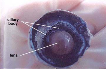
On the anterior hemisphere of the eye, there is the semi-fluid vitreous humor that fills the central cavity of the eye and the aqueous humor, which is found behind the cornea.
We scoped out the vitreous humor and the lens, this reveals the lens, ciliary body, and suspensory ligaments. The normal lens is convex shaped and somewhat elastic. It is held in place by the suspensory ligaments that in turn join with the smooth muscle containing ciliary body. We then removed the lens by pulling it free from its attachments. The lens was circular and felt kind of like uncooked tapioca.


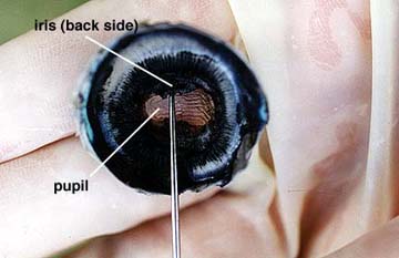
After removing the vitreous humor and the lens, we could now see an opening that allows light to enter the eye, the pupil, which is located in the center of the black iris. A sheep has an oblong- shaped pupil, while a human has a circular pupil. We then removed a thin filament protein covering the pupil on the front side of the hemisphere, called the cornea.


Wednesday, April 13, 2016

The Clay Brain

Today in class we made 2-dimensional clay brains out of playdoh. We used different colors for the different parts of the brain. Here are the pictures:


Right Cerebral Hemisphere:

Left Hemisphere along Sagittal Plane: 


Tuesday, April 12, 2016

"The Women with a hole in her brain"

We started a new unit in class today, the Nervous System, and we read an article relating to this subject. There is this women who hadn't realized till she was 24 that she did not have a cerebellum, the part of the brain responsible for body control and motion memory. She couldn't walk until she was seven and her speech only become intelligible at the age of six. Cerebrospinal fluid, which cushions the brain and provides defence against disease, took the place of the cerebellum. Problems in the cerebellum can lead to severe mental impairment, movement disorders, epilepsy or a potentially fatal build-up of fluid in the brain. However, in this woman, the missing cerebellum resulted in only mild to moderate motor deficiency, and mild speech problems such as slightly slurred pronunciation.

Now if we were to imagine a situation where a person did not have a thalamus or a damaged thalamus, would it is be possible for the person to survive or much less carry out daily functions? Could it have gone unnoticeable for a long time like the woman above? The thalamus is located towards the middle of the brain, above the brainstem and below cerebrum. It is responsible for sorting out of the data received via the spinal cord and direct it towards the corresponding part of the brain. The thalamus serves a gatekeeper that assures that messages are prioritized and regulated, some even call it the center of our self-identity. Without the benefit of the thalamus, a person is at the mercy of the countless messages flooding the brain, and cannot prioritize. Injury to the thalamus can produce distortions of sensory perception, which we see in many forms of brain dysfunction, but particularly in cerebral palsy and autism.