Tuesday, May 10, 2016

Reflexes Lab Analysis

Today in class we tested out our reflexes. Reflexes are rapid, predictable, and involuntary responses to stimuli. A reflex arc us a pathway to nerve impulses, they do not go to the brain.

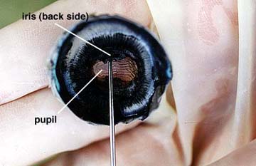
1. The Photopupillary Reflex
The smooth muscles of the iris function to control the size of the pupil. For example, the intensity of light entering the eye increases, the photopupillary relfex is triggered, and the cilliary body of the iris is stimulated to contract. As a result, the size of the pupil decreases, and the less light enters the eye.
For me, the reflex happened after I closed my eye for two minutes and then exposed it to bright light, which made my pupil decrease in size.
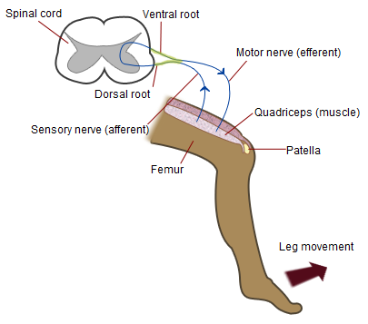
2. Knee Jerk Reflex
This reflex is called a monosynaptic reflex because there is only one synapse in the circuit needed to complete the reflex. A tap below the knee causes the thigh muscles to stretch. Information is then sent to the spinal cord. After one synapse in the ventral horn of the spinal cord, the information is sent back out the muscle and there you have the reflex.
For me, the reflex worked, even when I was trying to prevent it. My knee jerked forward. After doing 50 squats, I was very fatigued and the patellar reflex didn't work because I used all my ATP at my knee and the tap caused no muscle contraction.

3. Blink Reflex
The Blink reflex is designed to protect our eyes. Eyes are very fragile and cannot be replaces so it is very important to protect them, which this reflex does. If eyelids get hurt, they can repair themselves but our eyes cannot.
I blinked even with a thin transparent barrier when a cotton ball was thrown at me.

4. Babe, what is your sign?
In adults, a typical response to the plantar reflex, which is when one person strokes another person's sole of their feet from heel to base of the big toe, is that the toes will flex and move closer together. This happened for me. However if there is nerve damage one might show Babinski's sign, where the toes spread apart and upward. In newborn, Babinski's sign should always be seen since their nervous system is not completely  myelinated yet.


5. For the last reflex test, we are asked to catch a ruler dropped at unknown time, to determine the rate at which our relfexes work. For me I was able to catch the ruler at an average time of 0.28 seconds without texting and with texting at an average time of 0.18 seconds.
My reaction time was faster when texting because I already got practice catching the ruler and was more alert when catching the ruler. Although this shouldn't be the case because we perform tasks less efficiently when multitasking because our brains cannot multitask. It can only switch between tasks really quickly.

Here is our class's data:

Thursday, May 5, 2016

Your Brain Map


Cerebral Cortex
1.     What do the frontal lobes do?
  Your frontal lobes are your central-command center. The frontal lobe controls personality, but it's also where the problem solving, memory, language, judgement and impulse control and other behavioral traits occur. 


2.     What is the relationship between selective attention and learning?
S      Selective attention makes your working memory capacity greater as it allows you to prioritize information, often-used information over unimportant, little-used information. Learning is a process by which you engage with the subject in different ways so your brain can attain more important information. 


3.     What is the last part of your brain to develop and what can you do to prevent it from deteriorating?
 Your frontal lobe is the last part of our brains to develop. You can prevent it from deterioration by engaging with the environment, focusing on your five senses, and by transforming your ideas so that it can be developed into bigger ideas that stick to your brain for longer.

4.     What does the neo cortex do?
 The neocortex helps you to navigate your body by governing your senses, spatial awareness and motor skills.

5.     What is the role of the pre frontal cortex?
 Your prefrontal cortex controls your personality including how you behave in social situations. It also organizes your thoughts and actions to match cohesively with your internal goals, and the determination to study for long-hours.

6.     What do we know about the pre frontal cortex’s relationship with multitasking?
 The prefrontal cortex actually has a "central bottleneck" which is there to prevent you from doing more than two major things at one time. Your brain doesn't really multitask so much as so jump from one task to another with speed, which might make the quality of each task inferior.

7.     Which part of the brain is associated with speech and language development?  Give an interesting fact about this region.
 Broca's area is responsible for speech and language development. Broca's area is also associated with learning a second language.

8.     Which part of your brain is responsible for thinking the following: “Is it hot in here or is it just me?”
        The somatosensory cortex

9.     What does your visual cortex do for you?
 It helps differentiate colors and distinguish complex things such as faces

10.  State three interesting or significant facts about your occipital lobe.
 1.) It collects and categorizes visual data and routes it to other parts of your brain for identification and storage.
2. ) This part of the brain also deals with your imagination and allows you to process short and long-term memories.
3.) You can engage your occipital lobe by using your imagination to "visualize" a task over an over again, like a surgeon can mentally practice an operation, which will improve your practice in real life.

11.  What would happen if your temporal lobes were damaged?
 Without your temporal lobes, you would be unable to visualize memories, or comprehend language, process emotions and derive meanings. You would forget where you parked your car or where your house is.

12.  What is your “fast brain” and what does it do?
 The parietal lobes can quickly and efficiently process all the information it is getting from other sources, it deals with senses and navigation of your body.

Neuron
13.  State 3 things that you could do that would influence your synapses, and have a positive affect on your life and health.
 Exercising and a healthy diet will positively affect your life and synapses, as omega fatty acids provide good brain power and improve synapse function. Socializing is also proven to help fire more synapses.

14.  What is the relationship between multi-sensory or multi-modal learning and your dendrites?
 Multi-sensory or multi-modal simulation in dendrites lead to greater retention because the information is stored in more than one place.

15.  How does “big picture thinking” and mnemonics affect dendrites and/or learning?
 Once your brain sees the "big picture" it breaks down the information into smaller sections.  Mnemonics helps because it is an easy way to remember information due to its tangibility compared to abstract facts.

16.  Describe a neurotransmitter that you feel is very important.  Justify your reasoning.
 Dopamine is a natural neurotransmitter that makes you feel happy and it makes you want to try new things, which is an incentive for your brain to gain information.

Limbic System
17.  What does the corpus callosum do?
 It communicates between the right and left hemispheres. It is responsible for oral language skills and reading.


18.  What is the relationship between music and the corpus callosum?
 Music helps strengthen the connection between the two hemispheres, thereby making the corpus callosum stronger.

19.  Why is the thalamus important?
 It is important because it not only relays information to the cortex and is responsible for processing information but because it also is responsible for retrieving memories.


Relate and Review

Summarize what you learned from this tutorial.  Relate what you learned to your everyday life and how you can make it better.  Use at least 5 of the bolded words from the questions.  5-sentence minimum. You may use the back of this if needed.

The brain is composed of many different sections that each deal with a different task and situation, something functions of each part of the brain overlaps. The right and left hemispheres is connected by a corpus callosum, which can be strengthened through music. Another part of the brain, the visual cortex appears to be linked to the occipital lobe in that both provide the function of allowing us to see. Lastly, the temporal lobe proves to an important part of daily life as it helps me visualize memories. The temporal lobe is responsible for memory long-term and some short term. Neurons help us to take in information felt b our five senses and relay it to our brain, through neurotransmitters, that are chemical hormones made by our body.


Saturday, April 30, 2016

Sheep Brain Dissection

Today in class, we dissected a brain. We only made three cuts but we identified most of the important structures of the brain, by using pins.
 Anterior (White) and Posterior (Black)
Cerebrum (yellow), Cerebellum (green), and Brain Stem (Red)


First we observed the external structure of the brain, starting with the meninges, the a shiny membrane substance surrounding the brain. After removing the meninges, we identified the anterior and posterior sides of the brain. Then we identified the cerebrum, cerebellum, and brain stem.


Cerebrum
is divided into four lobes and is responsible for body orientation, processing sensory input, cognition, memory, voluntary motor action, visual reception and association, auditory reception and interpretation, and expression powers.
Cerebellum
is responsible for motor control and coordination.
Brain Stem
performs necessary functions, like heart rate and breathing

Myelin is a mixture of proteins and phospholipids forming a whitish insulating sheath around many nerve fibers, increasing the speed at which impulses are conducted. 


Then we cut the brain longitudinally (like a hot dog) so that we could observe the medial plane of the brain. From this view of the brain we could see the white and grey matter of the brain. 

 Thalamus (yellow), Midbrain (blue), Medulla Oblongata(Red), Pons (Green)


 Hypothalamus (Blue)


Corpus Callosum (Red)                                                                                          Optic Nerve (Green)


Thalamus
works to correlate several important processes, including consciousness, sleep, and sensory interpretation.
Optic Nerve
transfer visual information from the retina to the vision centers of the brain via electrical impulses. The optic nerve is made of ganglionic cells or nerve cells.
Medulla Oblongata
regulate breathing, heart and blood vessel function, digestion, sneezing, and swallowing. This part of the brain is a center for respiration and circulation.
Pons
a bridge between various parts of the nervous system, including the cerebellum and cerebrum
Midbrain
is a portion of the central nervous system associated with vision, hearing, motor control, sleep/wake, arousal (alertness), and temperature regulation.
Corpus Callosum
Large bundle of neuron fibers (myelinated axons or white matter) connecting 2 hemispheres
Hypothalamus
Maintains homeostasis and links the nervous system to the endocrine system

Then we made a cross sectional cut of the cerebrum (like a hamburger).

White and Gray Matter


Wednesday, April 27, 2016

Sheep Eye Dissection

Today in class, my partner and I dissected a sheep eye to better understand the structure and physiology of the eye. A sheep eye is a little bit different from the human eye, for example, a sheep's pupil is oblong shaped, while the human eye is circular. Additionally, the sheep eye has a glittery layer behind the retina called the tapetum lucidum, which helps sheep see in the dark. Humans don't need this because we don't have night-vision.

The eye untouched:


We started the dissection by cutting away the fatty tissue, the muscle, and eye-lid from both the front and rear surfaces of the eye. Then we were left with an eye that looks like this:


Then we made an incision down the midway of the sclera between the cornea and the optic nerve.
This cut the eye in two anterior and posterior hemispheres. Each side looked like this:


On the posterior (back) hemisphere of the eye there is the retina when peeled to the side reveals a black underlying choroid coat. The retina remains attached in one place called the blind spot. Then we scraped away the choroid coat, which revealed a blue glittery layer called the tapetum lucidum, which, as I have said above, is not found in the human eye because. It reflect light onto the retina which helps certain animals see in the dark.


On the anterior hemisphere of the eye, there is the semi-fluid vitreous humor that fills the central cavity of the eye and the aqueous humor, which is found behind the cornea.
We scoped out the vitreous humor and the lens, this reveals the lens, ciliary body, and suspensory ligaments. The normal lens is convex shaped and somewhat elastic. It is held in place by the suspensory ligaments that in turn join with the smooth muscle containing ciliary body. We then removed the lens by pulling it free from its attachments. The lens was circular and felt kind of like uncooked tapioca.


After removing the vitreous humor and the lens, we could now see an opening that allows light to enter the eye, the pupil, which is located in the center of the black iris. A sheep has an oblong- shaped pupil, while a human has a circular pupil. We then removed a thin filament protein covering the pupil on the front side of the hemisphere, called the cornea.


Wednesday, April 13, 2016

The Clay Brain

Today in class we made 2-dimensional clay brains out of playdoh. We used different colors for the different parts of the brain. Here are the pictures:


Right Cerebral Hemisphere:

Left Hemisphere along Sagittal Plane: 


Tuesday, April 12, 2016

"The Women with a hole in her brain"

We started a new unit in class today, the Nervous System, and we read an article relating to this subject. There is this women who hadn't realized till she was 24 that she did not have a cerebellum, the part of the brain responsible for body control and motion memory. She couldn't walk until she was seven and her speech only become intelligible at the age of six. Cerebrospinal fluid, which cushions the brain and provides defence against disease, took the place of the cerebellum. Problems in the cerebellum can lead to severe mental impairment, movement disorders, epilepsy or a potentially fatal build-up of fluid in the brain. However, in this woman, the missing cerebellum resulted in only mild to moderate motor deficiency, and mild speech problems such as slightly slurred pronunciation.

Now if we were to imagine a situation where a person did not have a thalamus or a damaged thalamus, would it is be possible for the person to survive or much less carry out daily functions? Could it have gone unnoticeable for a long time like the woman above? The thalamus is located towards the middle of the brain, above the brainstem and below cerebrum. It is responsible for sorting out of the data received via the spinal cord and direct it towards the corresponding part of the brain. The thalamus serves a gatekeeper that assures that messages are prioritized and regulated, some even call it the center of our self-identity. Without the benefit of the thalamus, a person is at the mercy of the countless messages flooding the brain, and cannot prioritize. Injury to the thalamus can produce distortions of sensory perception, which we see in many forms of brain dysfunction, but particularly in cerebral palsy and autism.